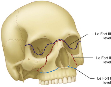
Direct impact to the central midface. Le Fort I most inferior.

Facial Fracture Management Handbook Lefort Fractures Iowa Head And Neck Protocols
The oral cavity should be assessed for mucosal lacerations ecchymosis and the presence of.

. Therefore these fractures are also called pyramidal fractures. Transverse fracture through the maxillary sinuses lower nasal septum pterygoid plates. -The frequency of Le Fort types is in the order of type II type I.
Patients with Le Fort II injuries are often admitted to hospital unconscious and intubated. If significant these orbital fractures may be treated in the same way as other orbital fractures in this location. This is the highest level LeFort fracture and essentially separates the maxilla from the skull base.
A simple fracture is one in which there is no contact of the broken bone with the outer air ie the overlying tissues are intact. Introduction Le Fort fractures account for 10-20 of all facial fractures. Le Fort type III fractures are caused by impact to the nasal bridge and upper part of the maxilla.
A Le Fort III fracture involves fractures of the internal orbit. Le Fort fractures account for 10 to 20 of all facial fractures. Le Fort fracture is most likely present anterolateral margins of nasal fossa above maxillary alveolar ridge intact.
Other causes include assaults and falls. With seatbelt laws and the increased use of airbags by auto manufacturers the overall incidence of midface fractures has decreased. Le Fort I.
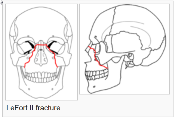
Pyramidal fracture apex of pyramid is nasofrontal suture Central midface and maxilla are mobilized independently from facial skeleton and cranial base. There are three main types of Le Fort fractures but there may be individual variations. The Le Fort II fracture extends posteriorly to the pterygoid plates at the base of the skull.
Le Fort III is excluded. Spans the body of the nasal septum. Direct horizontal impact to the upper jaw.
LeFort III fractures result in craniofacial disjunction. They result from exposure to a considerable amount of force. Maxillary occlusion bearing segment closed mouth with maxillary and mandibular approximation 2.
If the nasal complex moves along with the maxilla a LeFort II fracture is likely. The Le Fort II fracture is also referred to as a pyramidal fracture. Superior to the maxillary alveolar process.
Motor vehicle accidents are the predominant cause. Common etiologies include assault facial trauma in contact sports motor vehicle accidents MVA or falls from significant heights. The hallmark of Lefort fractures is traumatic pterygomaxillary separation which signifies fractures between the pterygoid plates horseshoe shaped bony protuberances which extend.
Le Fort II fracture is also called the pyramidal fracture pattern which is identified by the separation of nasomaxillary complex. Nasal septum lateral nasal walls lateral maxillary sinus wall and pterygoid plates are involved in these kinds of fractures. Rene Le Fort in 1901.
First these injuries often have multiple segments in which case comminution and compression of the maxilla may follow efforts to pull the maxilla en bloc. Describe the LeFort II fracture. Le Fort I is excluded inferior orbital rims fractured.
Le Fort II is likely present and zygomatic arches intact. The pterygoid plate involved in ALL Le Fort fractures The maxilla. Click here for further details on the reconstruction of combined medial wall and orbital floor fractures.
Pterygoid processes fractured in this case. These fractures are associated with malocclusion and dental fractures. Circumzygomatic suspension wiring of Le Fort II fractures has been described.
Oblique fracture crossing zygomaticomaxillary suture inferior orbital rim nasal bridge. 1 The Le Fort II fracture crosses the nasal bones on the ascending process of the maxilla and lacrimal bone and crosses the orbital rim. While this method may be effective for clean true Le Fort II fractures it is discouraged for 3 main reasons.
The fracture extends above the upper jaw maxilla. Le Fort type II fractures result from a force to lower or mid maxilla. A Le Fort fracture of the skull is a classic transfacial fracture of the midface involving the maxillary bone and surrounding structures in either a horizontal pyramidal or transverse direction.
The face is made up of several bones and a type II fracture will involve several of the following bones. Described by Dr. Initially described in 1901 by French surgeon René Le Fort 1869-1951 LeFort fractures represent a group of midface fractures that occur following blunt trauma and follow areas of structural weakness.
The mandibular and maxillary fixation is performed before fixation of the upper mid face fractures along the four maxillary buttresses bilateral nasomaxillary and zygomaticomaxillary buttresses. Le Fort II. Nasal and lacrimal bones nasofrontal suture infraorbital rims and pterygoid plates are involved in this.
May involve pterygoid plates of the sphenoid bone. Trans-maxillary fracture also known as Guerin fracture Bony disruption involves. The fracture extends from the lower part of one cheek below the eye across the bridge of.
To classify this Le Fort fracture look at the following four facial segments. Fracture breaking of a bone. Special attention has to be paid to foreign bodies such as teeth or tooth fragments obstructing the.
At the level of the infraorbital rim and nasofrontal junction. LeFort II fractures transect the nasal bones medial-anterior orbital walls orbital floor inferior orbital rims and finally transversely fracture the posterior maxilla and pterygoid plates. -Le Fort and maxillary fractures accounted for 255 of 663 facial fractures recently reported from a level 1 trauma center 3.
Ecchymosis facial edema subcutaneous hematomas and epistaxis are often seen in maxillary fractures. If all of these bones are broken it forms a pyramidal structure around the nose. It commonly extends from the pterygoid plate through the maxilla through the nasal orbital ethmoid area and nasofrontal bone.
Closed Treatment For Le Fort Ii


0 Comments